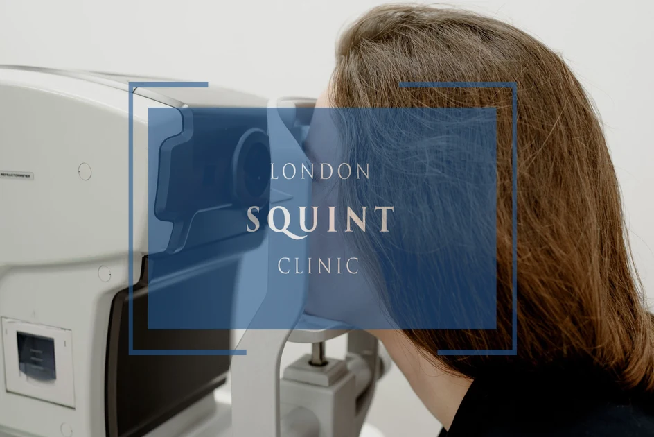
Brain Tumor Double Vision: Post-Surgery Eye Treatment London

Why Choose Private Squint Surgery?
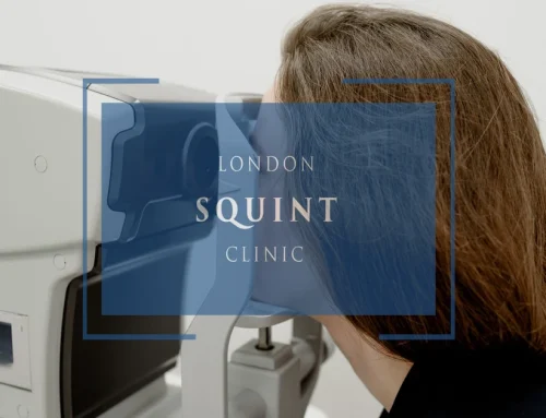
In the UK, squint surgery may be performed via the NHS, through insurance, or as self-pay. NHS treatment is free, but waiting times can be long and surgery is often performed by trainees under supervision. Many operations are carried out by surgeons who mainly specialise in children’s squint rather than adult complex cases.
With insurance, fees are standardised — meaning some leading specialists choose not to participate. Self-pay allows you to choose your surgeon directly and prioritise experience, specialisation, and access.
Many centres quote only a surgical fee. Hospital costs, anaesthetic fees and follow-ups are frequently additional. At London Squint Clinic, everything is included in one transparent package.
Our Complete Package – £10,000
- ✔ Advanced surgery by Mr Ali (one or both eyes)
- ✔ Adjustable sutures where clinically indicated
- ✔ Detailed orthoptic planning
- ✔ All hospital & anaesthetic fees included
- ✔ Post-operative medication
- ✔ Two video follow-ups
- ✔ Face-to-face review appointment
What Makes Us Different
- ✔ 100% focused on adult squint & double vision surgery
- ✔ >95% audited success rate
- ✔ Free re-treatment at 3 months if worse (extremely rare)
- ✔ 24/7 direct WhatsApp access to your surgeon during recovery
- ✔ Optional well-being session & pre-op reassurance call
Initial consultation: from £150
Surgery typically within 4 weeks. No referral required. Self-pay only.
Brain Tumor Double Vision
- Double vision (diplopia) after brain tumor surgery often results from disruption to neural pathways controlling eye movement, particularly when tumors are located near the brainstem, cerebellum, or cranial nerves.
- Recovery prognosis varies significantly—temporary factors like swelling may resolve within 3-6 months, while direct nerve damage may require longer recovery periods or specialized intervention.
- Treatment options range from non-invasive approaches (prism therapy, Botox injections) to advanced strabismus surgery with adjustable sutures for persistent cases.
- Comprehensive rehabilitation strategies including orthoptic therapy, visual field adaptation, and functional vision training are essential for maximizing recovery outcomes.
- A multidisciplinary approach involving neuro-ophthalmologists, neurosurgeons, and rehabilitation specialists provides the most effective management of post-neurosurgical vision complications.
Table of Contents
- Understanding Double Vision After Brain Tumor Surgery
- How Brain Tumors Affect Vision and Eye Movement
- Common Eye Complications Following Neurosurgery
- Will Double Vision From Brain Tumors Improve Over Time?
- Specialized Treatment Options for Post-Surgery Diplopia
- London’s Expert Approach to Neurosurgical Vision Recovery
- Rehabilitation Strategies for Restoring Normal Vision
Understanding Double Vision After Brain Tumor Surgery
Double vision (diplopia) is a common and distressing visual complication that can occur following brain tumor surgery. This condition manifests when the brain receives two images of a single object, resulting in overlapping or adjacent visual perceptions. For patients who have undergone neurosurgical procedures, this symptom can significantly impact daily functioning and quality of life.
Brain tumor double vision typically develops due to disruption of the complex neural pathways that control eye movement and coordination. During surgical removal of brain tumors, particularly those located near the brainstem, cerebellum, or cranial nerve pathways, the delicate mechanisms responsible for synchronised eye movements may become compromised. This disruption can occur through direct surgical manipulation, postoperative swelling, or changes in intracranial pressure.
Patients experiencing post-brain surgery diplopia often report symptoms ranging from subtle image separation to complete doubling of their visual field. This visual disturbance may be constant or intermittent, worsen with fatigue, and vary depending on the direction of gaze. The condition can cause significant challenges with reading, driving, spatial orientation, and even basic mobility, often necessitating specialised neuro-ophthalmological intervention.
How Brain Tumors Affect Vision and Eye Movement
Brain tumors can impact vision and ocular motility through various mechanisms, depending on their location, size, and growth pattern. Tumors affecting the visual pathway, cranial nerves, brainstem, or cerebellum are particularly likely to cause visual disturbances, including double vision. Understanding these mechanisms is crucial for effective management and treatment planning.
When tumors develop near cranial nerves III (oculomotor), IV (trochlear), or VI (abducens), they can directly impair the nerve’s function through compression or infiltration. These cranial nerves control the extraocular muscles responsible for eye movement, and their dysfunction results in misalignment of the visual axes—a condition known as strabismus—leading to diplopia. For instance, a tumor affecting the sixth cranial nerve may cause an inward deviation of the eye (esotropia), while fourth nerve involvement typically results in vertical misalignment.
Tumors in the posterior fossa, including those affecting the cerebellum or brainstem, can disrupt the neural integration centres that coordinate binocular vision. Additionally, increased intracranial pressure from brain tumors can cause papilloedema (swelling of the optic disc), potentially leading to visual field defects and, in some cases, contributing to diplopia through mechanical effects on cranial nerves.
The relationship between tumor location and specific visual symptoms provides valuable diagnostic information. For example, tumors in the cavernous sinus often produce a characteristic pattern of ocular motor nerve palsies, while those affecting the cerebellopontine angle may cause a combination of hearing loss and diplopia. This neuroanatomical correlation helps specialists at London Squint Clinic develop targeted treatment approaches for each patient’s unique presentation.
Common Eye Complications Following Neurosurgery
Neurosurgical procedures, while often life-saving, can result in various ocular complications that require specialised management. Double vision represents just one of several potential visual disturbances that may emerge following brain tumor surgery. Understanding these complications is essential for comprehensive post-operative care and rehabilitation planning.
Cranial nerve palsies constitute a significant category of post-neurosurgery eye complications. Third nerve palsy may cause ptosis (drooping eyelid), pupillary abnormalities, and limited eye movement in multiple directions. Fourth nerve palsy typically results in vertical diplopia that worsens when looking down or tilting the head. Sixth nerve palsy causes horizontal double vision that increases when looking toward the affected side. These palsies may result from direct surgical trauma, traction during tumor removal, or postoperative swelling affecting the nerve pathways.
Nystagmus, characterised by involuntary rhythmic eye movements, can develop following surgery near the cerebellum or brainstem. This condition often exacerbates visual disturbances and may contribute to dizziness and balance problems. Similarly, internuclear ophthalmoplegia—a disorder affecting coordinated eye movements—can occur after procedures involving the brainstem’s medial longitudinal fasciculus.
Visual field defects represent another category of post-neurosurgical complications, particularly following operations near the optic chiasm, optic radiations, or visual cortex. These deficits may range from subtle peripheral vision loss to hemianopia (blindness in half the visual field). Unlike diplopia, which involves seeing two images, field defects result in areas of non-vision within the normal visual range.
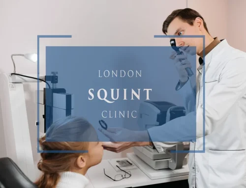
At London Squint Clinic, we conduct comprehensive neuro-ophthalmological assessments to identify the specific nature of post-neurosurgery visual complications, enabling targeted treatment approaches for each patient’s unique presentation.
Will Double Vision From Brain Tumors Improve Over Time?
The prognosis for double vision following brain tumor surgery varies considerably depending on multiple factors, including the underlying cause, extent of neural damage, patient age, and overall health status. Many patients and their families understandably seek clarity on whether their diplopia will resolve spontaneously or require intervention.
In cases where double vision results primarily from temporary factors such as postoperative swelling, inflammation, or mild nerve compression, significant improvement often occurs naturally within 3-6 months as these conditions resolve. This spontaneous recovery is particularly common in younger patients with good overall health and when the cranial nerves remain structurally intact. During this recovery period, temporary measures such as occlusion therapy (patching) or prism glasses may provide symptomatic relief.
However, when diplopia stems from direct damage to cranial nerves or their nuclei during tumor resection, the prognosis becomes more guarded. Partial recovery may still occur through neural adaptation and potential nerve regeneration, but this process typically takes longer—often 6-12 months—and may not result in complete resolution. In such cases, early intervention with specialised treatments can significantly improve functional outcomes and quality of life.
At London Squint Clinic, we carefully monitor patients with post-brain surgery diplopia through regular assessments to track recovery progress. This vigilant approach allows us to determine the optimal timing for intervention, avoiding premature surgical correction while not delaying treatment unnecessarily. Our experience with complex adult strabismus cases informs our prognostic assessments, enabling us to provide patients with realistic expectations regarding their visual recovery journey.
Specialized Treatment Options for Post-Surgery Diplopia
Managing double vision after brain tumor surgery requires a multidisciplinary approach tailored to each patient’s specific visual dysfunction. At London Squint Clinic, we offer comprehensive treatment options ranging from non-invasive therapies to advanced surgical interventions for persistent diplopia.
Prism therapy represents a valuable non-surgical option for many patients with post-neurosurgical diplopia. Fresnel prisms (temporary press-on prisms) or ground-in prisms incorporated into spectacle lenses can realign images on the retina, effectively eliminating double vision in primary gaze positions. This approach is particularly beneficial during the waiting period for spontaneous recovery or as a long-term solution for patients with stable, mild-to-moderate misalignment.
Botulinum toxin (Botox) injections offer another non-surgical intervention that can be highly effective for certain types of post-brain surgery strabismus. By temporarily weakening specific extraocular muscles, these injections can reduce misalignment and alleviate diplopia. They are especially useful in cases where the final outcome remains uncertain, as they provide temporary relief without permanent changes to ocular anatomy.
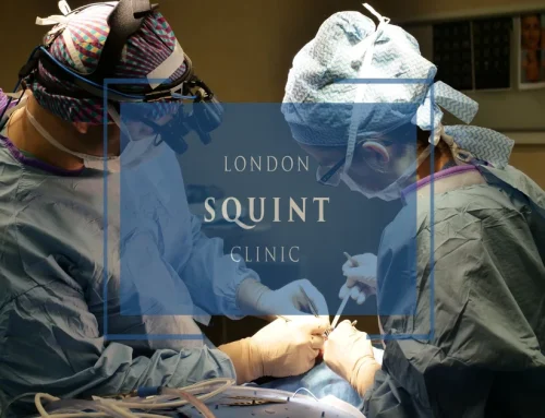
For patients with persistent diplopia that doesn’t respond adequately to conservative measures, strabismus surgery provides a definitive solution. This procedure involves precisely adjusting the tension of the extraocular muscles to realign the visual axes. Modern surgical techniques allow for adjustable sutures, enabling fine-tuning of alignment in the immediate postoperative period to optimise results. Our surgical approach for post-brain tumor diplopia is highly specialised, taking into account the unique challenges presented by neurological strabismus, including potential aberrant regeneration and restrictive components.
Vision therapy and orthoptic exercises complement these interventions by helping patients develop compensatory strategies, improve fusion ability, and expand the range of single binocular vision. This multifaceted treatment approach ensures that each aspect of post-neurosurgical diplopia is addressed, maximising functional visual recovery.
London’s Expert Approach to Neurosurgical Vision Recovery
London Squint Clinic has established itself as a centre of excellence for managing complex visual complications following brain tumor surgery. Our approach combines cutting-edge diagnostic technology, specialised surgical expertise, and comprehensive rehabilitation protocols specifically designed for neurological vision disorders.
The cornerstone of our approach is detailed neuro-ophthalmological assessment, which goes beyond standard eye examinations to evaluate the complex interplay between neurological function and ocular motility. Using advanced diagnostic tools such as video-oculography, high-resolution MRI with orbital protocols, and computerised diplopia mapping, we can precisely identify the underlying mechanisms of post-neurosurgical diplopia. This detailed assessment allows us to differentiate between various causes, including cranial nerve palsies, nuclear lesions, internuclear ophthalmoplegia, and skew deviation.
Our surgical planning for brain tumor-related diplopia incorporates sophisticated modelling of ocular biomechanics, accounting for the unique challenges presented by neurological strabismus. Unlike standard strabismus cases, post-neurosurgical diplopia often involves complex patterns of incomitant deviation, restrictive elements, and aberrant innervation that require specialised surgical approaches. Our surgeons have extensive experience with these challenging cases, employing techniques such as partial tendon transposition, adjustable suture surgery, and selective botulinum toxin augmentation to achieve optimal outcomes.
Collaboration with neurosurgeons, neurologists, and neuroradiologists forms an integral part of our approach, ensuring that treatment plans address both the ocular manifestations and any ongoing neurological issues. This multidisciplinary framework allows for comprehensive management of complex cases, particularly when visual symptoms may indicate tumor recurrence or other neurological complications requiring attention.
Patient education and support represent another crucial element of our approach. We recognise that visual disturbances following brain tumor surgery can be particularly distressing, occurring at a time when patients are already coping with significant health challenges. Our team provides detailed explanations, realistic prognosis discussions, and ongoing support throughout the recovery journey.
Rehabilitation Strategies for Restoring Normal Vision
Rehabilitation plays a crucial role in maximising visual recovery following brain tumor surgery. At London Squint Clinic, we implement comprehensive rehabilitation strategies that extend beyond addressing the mechanical aspects of eye misalignment to include neurological adaptation, visual processing, and functional vision training.
Orthoptic therapy forms the foundation of visual rehabilitation for many patients with post-neurosurgical diplopia. These specialised exercises aim to strengthen binocular vision, expand fusion ranges, and develop compensatory strategies for residual misalignment. For patients with partial cranial nerve recovery, carefully designed orthoptic protocols can enhance neural adaptation, potentially reducing the need for surgical intervention or improving surgical outcomes when procedures are necessary.
Visual field adaptation training benefits patients who experience both diplopia and visual field defects following brain tumor surgery. This approach uses systematic stimulation of affected visual field areas to improve awareness and functional use of remaining vision. Similarly, oculomotor training helps patients develop more efficient eye movement patterns to compensate for limitations in gaze range or stability.
Functional vision rehabilitation focuses on practical strategies for managing daily activities despite visual impairments. This includes techniques for reading, navigation, driving assessment, workplace adaptations, and technology utilisation. Our occupational therapists work closely with neuro-ophthalmology specialists to develop personalised approaches that address each patient’s specific visual challenges and life requirements.
Perceptual training addresses higher-level visual processing difficulties that may accompany brain tumor-related vision problems. These exercises target skills such as visual attention, spatial awareness, visual memory, and visual-motor integration—functions that may be affected by tumors or surgical interventions involving visual association areas of the brain.
Throughout the rehabilitation process, we employ objective outcome measures to track progress and refine treatment approaches. Regular assessments of diplopia fields, fusion ranges, contrast sensitivity, and functional vision questionnaires provide quantifiable data on recovery, helping patients recognise improvements that might otherwise seem subtle or incomplete. This evidence-based approach ensures that rehabilitation efforts remain targeted and effective, maximising each patient’s potential for visual recovery following brain tumor surgery.
Frequently Asked Questions
How long does double vision typically last after brain tumor surgery?
Double vision after brain tumor surgery typically improves within 3-6 months if caused by temporary factors like swelling or inflammation. When diplopia results from direct nerve damage, recovery may take 6-12 months or longer, and some patients may experience permanent double vision requiring intervention. Recovery timelines vary based on tumor location, surgical approach, patient age, and overall health status.
Can double vision after brain surgery be completely cured?
Yes, double vision after brain surgery can often be completely resolved through a combination of natural recovery, non-surgical interventions, and surgical correction if necessary. Complete resolution is most likely when diplopia stems from temporary causes like swelling. Even in cases with permanent nerve damage, treatments including prism glasses, botulinum toxin injections, or strabismus surgery can effectively eliminate double vision in primary gaze positions for most patients.
What treatments are available for double vision that doesn’t improve on its own?
Persistent double vision after brain tumor surgery can be treated with several options: prism glasses to realign images without surgery, botulinum toxin injections to temporarily weaken specific eye muscles, strabismus surgery to permanently realign the eyes, vision therapy to improve fusion ability, and compensatory techniques like partial occlusion. Treatment selection depends on the specific cause, stability of the condition, and patient preferences.
How can I tell if my post-surgery double vision is improving?
Improvement in post-surgery double vision may be noticed through: decreased separation between the double images, smaller range of gaze positions triggering diplopia, reduced need for compensatory head positioning, improved ability to fuse images with minimal assistance, less visual fatigue during daily activities, and better depth perception. Regular measurements by a neuro-ophthalmologist using diplopia charting can objectively document progress even when changes feel subtle.
Are there specific brain tumor locations that are more likely to cause double vision?
Brain tumors most likely to cause double vision are those affecting the brainstem, cerebellum, cavernous sinus, and cranial nerve pathways. Specifically, tumors near cranial nerves III (oculomotor), IV (trochlear), and VI (abducens) commonly cause diplopia. Pituitary tumors, acoustic neuromas, meningiomas at the skull base, and metastatic tumors in these regions have higher associations with double vision compared to tumors in other brain areas.
When should I consider surgery for double vision after brain tumor removal?
Surgery for double vision should be considered when: symptoms persist beyond 6-12 months without improvement, non-surgical treatments like prism glasses provide inadequate relief, the eye misalignment is stable for at least 6 months, double vision significantly impacts quality of life or safety, and neurological recovery has plateaued. Most specialists recommend waiting until the condition stabilizes to avoid unnecessary procedures, as premature intervention may lead to suboptimal results.
Can vision therapy help with double vision caused by brain tumors?
Yes, vision therapy can help with brain tumor-related double vision by improving fusion ability, expanding the range of single binocular vision, developing compensatory eye movement patterns, and training the brain to suppress the second image in certain gaze positions. While vision therapy alone may not resolve severe misalignments, it works effectively as part of a comprehensive treatment plan, especially for mild to moderate diplopia or as rehabilitation following surgical correction.
Related Posts
Find out if you are suitable for Double Vision Treatment
Find out if you could benefit from this life changing surgery by contacting us today
Our most popular procedures


Hello, I’m Nadeem Ali
I’m one of the few eye surgeons in the world with 100% focus on Squint and Double Vision Surgery.
I have 24 years of eye surgery experience, and worked for 13 years as a Consultant at London’s renowned Moorfields Eye Hospital.
In 2023, I left the NHS to focus fully on treating patients from across the world at the London Squint Clinic. You can read more about me here.
There’s lots of information on the website about: squint surgery, double vision surgery and our pricing.
The most rewarding part of my job is hearing patients tell me how squint or double vision surgery has changed their lives. You can hear these stories here.
Mr Nadeem Ali
MA MB BChir MRCOphth FRCSEd(Ophth)