Post-Retinal Surgery Double Vision: When Eye Operations Cause Diplopia

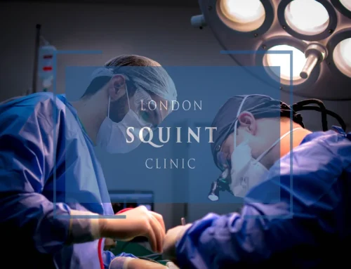
Why Choose Private Squint Surgery?
In the UK, squint surgery may be performed via the NHS, through insurance, or as self-pay. NHS treatment is free, but waiting times can be long and surgery is often performed by trainees under supervision. Many operations are carried out by surgeons who mainly specialise in children’s squint rather than adult complex cases.
With insurance, fees are standardised — meaning some leading specialists choose not to participate. Self-pay allows you to choose your surgeon directly and prioritise experience, specialisation, and access.
Many centres quote only a surgical fee. Hospital costs, anaesthetic fees and follow-ups are frequently additional. At London Squint Clinic, everything is included in one transparent package.
Our Complete Package – £10,000
- ✔ Advanced surgery by Mr Ali (one or both eyes)
- ✔ Adjustable sutures where clinically indicated
- ✔ Detailed orthoptic planning
- ✔ All hospital & anaesthetic fees included
- ✔ Post-operative medication
- ✔ Two video follow-ups
- ✔ Face-to-face review appointment
What Makes Us Different
- ✔ 100% focused on adult squint & double vision surgery
- ✔ >95% audited success rate
- ✔ Free re-treatment at 3 months if worse (extremely rare)
- ✔ 24/7 direct WhatsApp access to your surgeon during recovery
- ✔ Optional well-being session & pre-op reassurance call
Initial consultation: from £150
Surgery typically within 4 weeks. No referral required. Self-pay only.
Post-Retinal Surgery Double Vision
- Prevalence and Mechanisms: Double vision affects 5-25% of retinal surgery patients, commonly resulting from altered eye mechanics, inflammation, or extraocular muscle disruption.
- Procedure-Specific Risks: Vitrectomy (10-20% diplopia risk) and scleral buckling (5-25% risk) carry higher chances of alignment issues than less invasive procedures.
- Natural Resolution: 50-70% of cases improve or resolve completely within 3-6 months without intervention, particularly when caused by temporary factors like inflammation.
- Treatment Options: Management follows a stepwise approach from observation and prism therapy to orthoptic exercises and, if necessary, botulinum toxin injections or strabismus surgery.
- When to Seek Help: Consult a specialist if double vision persists beyond 4-6 weeks without improvement, suddenly worsens, or significantly impacts daily functioning.
Table of Contents
- Understanding Post-Retinal Surgery Diplopia: Causes and Mechanisms
- Common Types of Retinal Procedures That May Lead to Double Vision
- Why Does Double Vision Occur After Vitrectomy and Macular Surgery?
- Diagnosing Diplopia Following Retinal Surgical Interventions
- Treatment Options for Post-Retinal Surgery Double Vision
- Will My Double Vision After Eye Surgery Eventually Resolve?
- Prevention Strategies and Risk Factors for Surgical Diplopia
- When to Seek Specialized Care for Persistent Visual Disturbances
Understanding Post-Retinal Surgery Diplopia: Causes and Mechanisms
Post-retinal surgery diplopia refers to double vision that develops following procedures to repair or treat retinal conditions. This visual disturbance can be particularly distressing for patients who undergo retinal surgery expecting improved vision, only to find themselves struggling with a new visual challenge. The development of diplopia after retinal interventions is not uncommon, affecting approximately 5-25% of patients depending on the specific procedure performed.
The mechanisms behind post-retinal surgery diplopia are multifaceted. Most commonly, the condition stems from alterations to the eye’s mechanical properties during surgery. Retinal procedures often involve manipulation of ocular tissues, placement of silicone oil or gas bubbles, or the application of scleral buckles—all of which can affect the normal functioning of extraocular muscles. These muscles control eye movement and alignment, and when their function is compromised, the eyes may no longer work in perfect coordination.
Additionally, post-operative inflammation, scarring of orbital tissues, or direct trauma to the muscles during surgery can contribute to misalignment. In some cases, pre-existing minor eye alignment issues that were previously well-compensated may become symptomatic following the stress of surgery and anaesthesia. Understanding these mechanisms is crucial for both prevention and effective management of post-retinal surgery visual disturbances.
Common Types of Retinal Procedures That May Lead to Double Vision
Several retinal surgical interventions carry varying risks of post-operative diplopia. Vitrectomy, one of the most frequently performed retinal procedures, involves removing the vitreous gel from the eye to access the retina. This procedure is particularly associated with post-operative double vision, especially when combined with other interventions. Studies indicate that approximately 10-20% of patients may experience some degree of diplopia following vitrectomy.
Scleral buckling procedures, used primarily for retinal detachment repair, involve placing a silicone band around the eye to push the wall of the eye against the detached retina. This mechanical alteration can directly affect extraocular muscle function, with diplopia rates reported between 5-25%. The buckle may restrict normal eye movement or create mechanical imbalance between the eyes.
Macular hole and macular pucker surgeries, which often require meticulous work near the central retina, can also lead to diplopia. These procedures frequently involve gas bubble placement and face-down positioning during recovery, which may contribute to temporary or permanent changes in eye alignment.
Retinal detachment repair using pneumatic retinopexy (injection of a gas bubble) generally has lower rates of post-operative diplopia compared to more invasive procedures, but still carries some risk. Complex cases involving proliferative vitreoretinopathy or those requiring silicone oil tamponade tend to have higher rates of post-operative alignment issues due to the more extensive manipulation required and the prolonged presence of oil in the eye.
Why Does Double Vision Occur After Vitrectomy and Macular Surgery?
The development of diplopia following vitrectomy and macular surgery can be attributed to several specific mechanisms. During vitrectomy, the insertion of instruments through the sclera can potentially damage or irritate the extraocular muscles, particularly the inferior oblique or inferior rectus muscles. These muscles are responsible for eye movement and alignment, and any disruption to their function can result in misalignment and subsequent double vision.
Macular surgery often requires prolonged surgical manipulation near the macula, the central part of the retina responsible for detailed vision. This extended surgical time increases the risk of inflammation and swelling around the extraocular muscles. Additionally, the post-operative requirement for face-down positioning with gas bubble placement can create temporary imbalances in the visual system as the brain adapts to unusual visual input during the recovery period.
Another significant factor is the use of local anaesthesia. Retrobulbar or peribulbar injections used during these procedures can directly affect muscle function or cause localised tissue trauma. In some cases, the anaesthetic may temporarily paralyse certain extraocular muscles, leading to imbalance that persists even after the anaesthetic wears off.
Mechanical factors also play a crucial role. The placement of silicone oil or gas bubbles changes the weight and balance of the eye, potentially altering its position in the orbit. Similarly, post-operative scarring can restrict normal muscle movement, creating tension that pulls the eye out of alignment with its fellow eye. Understanding these specific mechanisms helps in both preventing and appropriately treating post-vitrectomy diplopia.
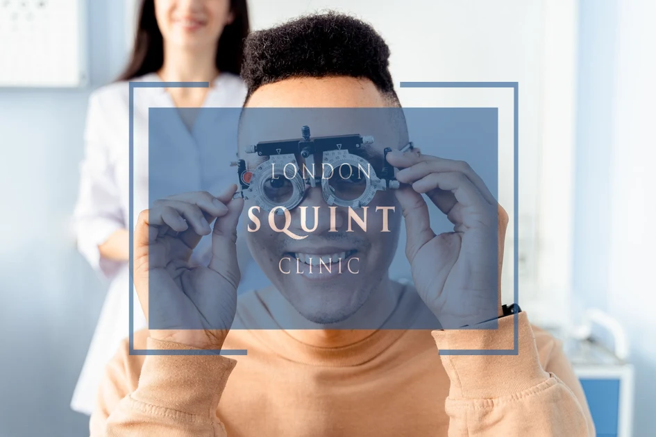
Diagnosing Diplopia Following Retinal Surgical Interventions
Accurate diagnosis of post-retinal surgery diplopia requires a comprehensive approach that begins with a detailed patient history. Ophthalmologists must determine when the double vision started in relation to surgery, whether it is constant or intermittent, and if it changes with gaze direction or head positioning. These temporal and situational factors provide crucial clues about the underlying mechanism.
Clinical examination typically includes several specific tests. The cover-uncover test assesses eye alignment by observing how each eye moves when the other is briefly covered. Prism measurements quantify the degree of misalignment in different gaze positions. The Hess screen or Lancaster red-green tests map the function of individual extraocular muscles, helping to identify which specific muscles may be affected.
Orthoptic assessment is particularly valuable, as orthoptists specialise in eye movement disorders and can perform detailed measurements of ocular motility. They can distinguish between comitant strabismus (where the degree of misalignment is the same in all directions of gaze) and incomitant strabismus (where the misalignment varies with gaze direction), which has important implications for treatment.
In complex cases, additional imaging may be necessary. Orbital MRI can reveal mechanical restrictions, muscle swelling, or other structural abnormalities that might explain persistent diplopia. CT scanning may be useful when suspecting orbital wall fractures or other bony abnormalities that could affect eye alignment. A thorough diagnostic approach ensures appropriate treatment planning and helps distinguish temporary from potentially permanent diplopia following retinal surgery.
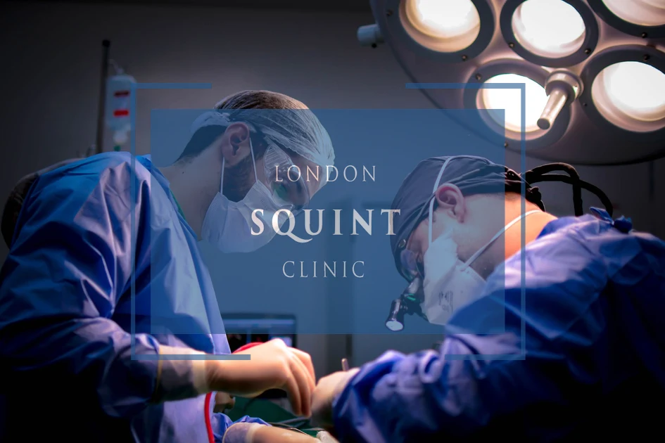
Treatment Options for Post-Retinal Surgery Double Vision
The management of post-retinal surgery diplopia follows a stepwise approach, beginning with conservative measures before progressing to more invasive interventions if necessary. Initially, a period of observation may be recommended, particularly in the early post-operative period, as some cases of diplopia resolve spontaneously as inflammation subsides and the eye heals.
Prism therapy represents a non-invasive first-line treatment option. Fresnel prisms, which are thin plastic sheets applied to spectacle lenses, can realign images and eliminate double vision without surgery. These prisms can be adjusted as the condition evolves and may provide either temporary relief during the healing process or a permanent solution for stable diplopia.
Orthoptic exercises may benefit selected patients, particularly those with small-angle deviations or those with convergence insufficiency following surgery. These exercises aim to strengthen eye muscle coordination and can improve fusion ability, helping the brain to combine the images from both eyes more effectively.
For persistent diplopia that doesn’t respond to conservative measures, strabismus surgery may be necessary. This involves adjusting the tension of the extraocular muscles to realign the eyes. The specific surgical approach depends on the pattern of misalignment, with options including muscle recession (weakening), resection (strengthening), or transposition procedures. In cases related to scleral buckle, removing or adjusting the buckle might be considered if the retina remains stable.
Botulinum toxin (Botox) injections offer a less invasive alternative to surgery for some patients. These injections temporarily weaken specific extraocular muscles, potentially allowing for realignment. While the effect is temporary (typically lasting 3-6 months), repeated injections can sometimes lead to more permanent improvement, or at least provide a therapeutic trial before committing to surgery.
Will My Double Vision After Eye Surgery Eventually Resolve?
The prognosis for post-retinal surgery diplopia varies considerably depending on several factors. Temporary diplopia, which resolves spontaneously, is relatively common following retinal procedures. Approximately 50-70% of cases show significant improvement or complete resolution within 3-6 months without specific intervention beyond the normal healing process. This is particularly true for diplopia related to post-operative inflammation, temporary muscle dysfunction due to anaesthesia, or adaptation to new visual conditions.
Several prognostic factors help predict the likelihood of spontaneous resolution. Diplopia that is intermittent rather than constant generally has a better prognosis. Similarly, double vision that improves steadily over the first few weeks after surgery typically continues to resolve completely. The magnitude of the deviation is also important—smaller deviations (less than 10 prism dioptres) are more likely to resolve spontaneously than larger ones.
The underlying mechanism significantly influences the timeline and likelihood of resolution. Diplopia caused by temporary factors such as gas bubble presence or post-operative inflammation typically resolves as these factors dissipate. In contrast, diplopia resulting from mechanical restrictions (such as scleral buckle placement) or direct muscle trauma may be more persistent.
For patients with diplopia that persists beyond 3-6 months, the condition is less likely to resolve spontaneously. However, even in these cases, appropriate interventions can successfully manage the double vision in the vast majority of patients. It’s important to maintain regular follow-up appointments with your ophthalmologist or strabismus specialist during this period to monitor progress and adjust treatment plans as needed.
Prevention Strategies and Risk Factors for Surgical Diplopia
Certain patient characteristics are associated with increased risk of developing post-retinal surgery diplopia. Pre-existing strabismus, even if previously asymptomatic, significantly increases the likelihood of post-operative alignment problems. Patients with a history of previous eye surgery, particularly those involving extraocular muscles, face elevated risk. Additionally, individuals with thyroid eye disease, myopia (near-sightedness), or advanced age may be more susceptible to post-surgical diplopia.
Surgical technique modifications can help minimise the risk of post-operative diplopia. Careful placement of sclerotomy sites during vitrectomy, avoiding the insertion areas of extraocular muscles, may reduce the likelihood of muscle damage. When scleral buckles are necessary, using segmental rather than encircling buckles where appropriate can limit restriction of eye movements. Minimising surgical manipulation and operating time may also reduce post-operative inflammation that contributes to diplopia.
Anaesthetic considerations play an important role in prevention. Sub-Tenon’s anaesthesia generally carries a lower risk of direct muscle trauma compared to retrobulbar injections. For patients identified as high-risk, consideration of general anaesthesia may be appropriate to avoid potential complications from local anaesthetic techniques.
Pre-operative assessment should include a thorough evaluation of ocular motility and alignment, particularly in patients with risk factors. Identifying pre-existing minor deviations allows surgeons to counsel patients appropriately about increased risk and potentially modify surgical approaches. Post-operatively, early recognition and management of diplopia can prevent the development of more persistent problems, making regular follow-up essential for high-risk patients.
When to Seek Specialized Care for Persistent Visual Disturbances
While some degree of visual disturbance is expected immediately following retinal surgery, certain symptoms warrant prompt medical attention. Patients should seek immediate care if they experience sudden onset of double vision accompanied by severe pain, significant redness, or dramatic reduction in vision. These symptoms could indicate serious complications such as infection, increased intraocular pressure, or retinal re-detachment rather than simple post-operative diplopia.
Timing is a crucial factor in determining when to seek specialized evaluation for diplopia. If double vision persists beyond 4-6 weeks after surgery without improvement, consultation with a strabismus specialist is advisable. Similarly, if initially improving diplopia suddenly worsens or changes in character, this may indicate evolving mechanical issues that require assessment.
The nature of the diplopia itself provides important clues about when specialized care is needed. Double vision that varies significantly with gaze direction (incomitant strabismus) or that worsens over time rather than improving suggests mechanical restrictions or muscle damage that may benefit from early intervention. Diplopia that significantly impacts daily functioning—interfering with reading, driving, or work activities—should prompt referral to a specialist regardless of the time elapsed since surgery.
When seeking specialized care, patients should ideally be referred to ophthalmologists with specific expertise in strabismus and diplopia management, particularly those familiar with post-retinal surgery complications. These specialists can offer comprehensive assessment including detailed measurements of ocular alignment in all gaze positions, sensory testing to evaluate binocular vision status, and development of tailored treatment plans that may include prismatic correction, specialized exercises, or surgical intervention when appropriate.
Remember that early intervention for persistent diplopia often leads to better outcomes, as prolonged double vision can lead to development of suppression or other adaptive mechanisms that may complicate later treatment. Most importantly, with appropriate specialized care, the vast majority of patients with post-retinal surgery diplopia can achieve comfortable single vision, whether through non-surgical or surgical means.
Frequently Asked Questions
How common is double vision after retinal surgery?
Double vision (diplopia) affects approximately 5-25% of patients following retinal surgery, with variation depending on the specific procedure. Vitrectomy procedures have diplopia rates of 10-20%, while scleral buckling procedures show rates between 5-25%. Complex cases involving silicone oil or extensive manipulation typically have higher incidence rates.
How long does double vision last after retinal surgery?
Approximately 50-70% of post-retinal surgery diplopia cases resolve spontaneously within 3-6 months. Temporary diplopia related to inflammation, anesthesia effects, or gas bubble presence typically improves as these factors resolve. Double vision that persists beyond 6 months is less likely to resolve without intervention but remains treatable with prism therapy, orthoptic exercises, or surgery.
Can a vitrectomy cause permanent double vision?
Yes, vitrectomy can cause permanent double vision in some cases, particularly when extraocular muscles are damaged during surgery or when significant scarring develops. However, even permanent diplopia is usually manageable with appropriate treatment. Risk factors for permanent diplopia include pre-existing strabismus, previous eye surgeries, and extensive surgical manipulation during the vitrectomy procedure.
What treatments are available for double vision after eye surgery?
Treatments for post-surgical diplopia include: 1) Observation during the initial healing period, 2) Prism therapy using Fresnel prisms applied to eyeglasses, 3) Orthoptic exercises to improve eye coordination, 4) Botulinum toxin injections to temporarily weaken specific eye muscles, and 5) Strabismus surgery to permanently realign the eyes. Treatment selection depends on the cause, severity, and duration of the diplopia.
Should I be concerned about double vision immediately after retinal surgery?
Some degree of double vision immediately after retinal surgery is relatively common and often temporary. However, you should contact your surgeon if the diplopia is accompanied by severe pain, significant redness, or dramatic vision reduction, as these may indicate complications requiring immediate attention. Otherwise, report persistent diplopia at your scheduled follow-up appointments.
Can double vision after retinal surgery be prevented?
While not all cases can be prevented, risk reduction strategies include: careful placement of surgical instruments to avoid extraocular muscles, using segmental rather than encircling buckles when possible, minimizing surgical manipulation and operating time, considering sub-Tenon’s anesthesia instead of retrobulbar injections, and thorough pre-operative assessment to identify patients with pre-existing alignment issues who may need modified approaches.
When should I see a specialist for double vision after my retinal procedure?
Seek specialized care if your double vision persists beyond 4-6 weeks without improvement, worsens after initial improvement, varies significantly with gaze direction, or substantially impacts daily activities like reading or driving. Ideally, consult an ophthalmologist with expertise in strabismus management who understands post-retinal surgery complications. Early intervention often leads to better outcomes for persistent diplopia.
Related Posts


Find out if you are suitable for Double Vision Treatment
Find out if you could benefit from this life changing surgery by contacting us today
Our most popular procedures


Hello, I’m Nadeem Ali
I’m one of the few eye surgeons in the world with 100% focus on Squint and Double Vision Surgery.
I have 24 years of eye surgery experience, and worked for 13 years as a Consultant at London’s renowned Moorfields Eye Hospital.
In 2023, I left the NHS to focus fully on treating patients from across the world at the London Squint Clinic. You can read more about me here.
There’s lots of information on the website about: squint surgery, double vision surgery and our pricing.
The most rewarding part of my job is hearing patients tell me how squint or double vision surgery has changed their lives. You can hear these stories here.
Mr Nadeem Ali
MA MB BChir MRCOphth FRCSEd(Ophth)