Double Vision Explained: Neurological Disorders That Might Be to Blame

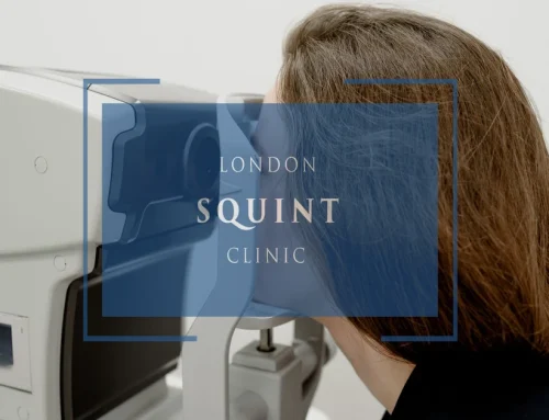
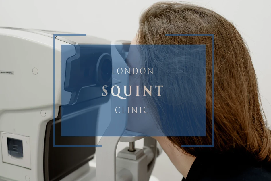
Why Choose Private Squint Surgery?
In the UK, squint surgery may be performed via the NHS, through insurance, or as self-pay. NHS treatment is free, but waiting times can be long and surgery is often performed by trainees under supervision. Many operations are carried out by surgeons who mainly specialise in children’s squint rather than adult complex cases.
With insurance, fees are standardised — meaning some leading specialists choose not to participate. Self-pay allows you to choose your surgeon directly and prioritise experience, specialisation, and access.
Many centres quote only a surgical fee. Hospital costs, anaesthetic fees and follow-ups are frequently additional. At London Squint Clinic, everything is included in one transparent package.
Our Complete Package – £10,000
- ✔ Advanced surgery by Mr Ali (one or both eyes)
- ✔ Adjustable sutures where clinically indicated
- ✔ Detailed orthoptic planning
- ✔ All hospital & anaesthetic fees included
- ✔ Post-operative medication
- ✔ Two video follow-ups
- ✔ Face-to-face review appointment
What Makes Us Different
- ✔ 100% focused on adult squint & double vision surgery
- ✔ >95% audited success rate
- ✔ Free re-treatment at 3 months if worse (extremely rare)
- ✔ 24/7 direct WhatsApp access to your surgeon during recovery
- ✔ Optional well-being session & pre-op reassurance call
Initial consultation: from £150
Surgery typically within 4 weeks. No referral required. Self-pay only.
Double Vision
- Diplopia (double vision) can be a warning sign of serious neurological conditions including stroke, multiple sclerosis, brain tumors, and raised intracranial pressure.
- Binocular diplopia, which resolves when either eye is covered, typically indicates a neurological issue affecting eye movement coordination or visual processing.
- Multiple sclerosis commonly causes diplopia through brainstem lesions, with approximately 40% of MS patients experiencing double vision during their disease course.
- The brainstem contains critical nuclei for eye movement control (cranial nerves III, IV, and VI), making it a key region where lesions frequently manifest as diplopia.
- Sudden double vision, especially when accompanied by headache, weakness, or coordination difficulties, requires urgent medical evaluation to rule out life-threatening conditions.
- Treatment approaches include addressing the underlying neurological condition while managing visual symptoms through prisms, occlusion, medications, or surgery in selected cases.
Table of Contents
- Understanding Diplopia: When Your Brain Sees Double
- Common Neurological Causes of Double Vision
- Multiple Sclerosis and Its Impact on Vision
- How Brainstem Lesions Affect Eye Movement Control
- Raised Intracranial Pressure: A Serious Vision Warning
- What Part of the Brain Causes Double Vision?
- Diagnosing the Root Cause of Neurological Diplopia
- Treatment Options for Neurologically-Induced Double Vision
Understanding Diplopia: When Your Brain Sees Double
Diplopia, commonly known as double vision, occurs when a person sees two images of a single object. This visual disturbance can be alarming and significantly impact daily functioning. While many associate double vision with eye problems, neurological disorders are frequently the underlying cause, particularly when the symptom appears suddenly.
Double vision can be categorised as either monocular (affecting one eye) or binocular (affecting both eyes). Monocular diplopia persists when one eye is covered and often stems from issues within the eye itself, such as cataracts or retinal problems. Binocular diplopia, which resolves when either eye is covered, typically indicates a neurological issue affecting the coordination of eye movements or the processing of visual information in the brain.
Sudden temporary double vision often signals a neurological event requiring urgent assessment. When diplopia appears abruptly, particularly in elderly patients or those with vascular risk factors, it may indicate serious conditions such as stroke, aneurysm, or increased intracranial pressure. Understanding whether double vision is neurological involves evaluating associated symptoms like headache, weakness, numbness, or coordination difficulties.
The brain’s complex visual system requires precise coordination between multiple cranial nerves, eye muscles, and neural pathways. When any component of this system is compromised by neurological disease, the result can be misalignment of the eyes and consequent double vision. This symptom should never be dismissed, as it may be the first warning sign of a significant neurological disorder.
Common Neurological Causes of Double Vision
Neurological diplopia can stem from various disorders affecting different parts of the visual pathway and brain. The most common cause of double vision from a neurological perspective is cranial nerve palsy, particularly involving the oculomotor (third), trochlear (fourth), or abducens (sixth) nerves that control eye movements. These palsies can result from microvascular damage due to diabetes or hypertension, particularly in older adults.
Myasthenia gravis, an autoimmune neuromuscular disorder, frequently presents with fluctuating diplopia that worsens with fatigue. The condition affects the neuromuscular junction, impairing the signals between nerves and eye muscles, resulting in weakness and misalignment.
Stroke remains a significant cause of sudden double vision, particularly when accompanied by other neurological deficits. Both ischaemic and haemorrhagic strokes affecting the brainstem or areas controlling eye movements can result in persistent diplopia. Similarly, transient ischaemic attacks (TIAs) may cause temporary double vision that resolves within 24 hours but serves as a critical warning sign.
Brain tumours, particularly those affecting the brainstem, cerebellum, or cranial nerves, can gradually compress structures responsible for coordinating eye movements. This compression often leads to progressive diplopia that may initially be intermittent before becoming constant.
Traumatic brain injuries frequently disrupt the delicate neural networks controlling binocular vision, resulting in persistent double vision. Even seemingly minor head trauma can damage cranial nerves or their nuclei, leading to diplopia that may resolve slowly or become permanent.
Vertical double vision specifically often indicates a fourth nerve palsy or skew deviation from brainstem dysfunction. Understanding these various neurological causes is essential for proper diagnosis and management of diplopia.
Multiple Sclerosis and Its Impact on Vision
Multiple sclerosis (MS) frequently affects the visual system, with diplopia being a common presenting symptom in many patients. This autoimmune condition causes demyelination—damage to the protective covering of nerve fibres—throughout the central nervous system, including areas responsible for eye movement control and visual processing.
In MS, double vision typically results from lesions in the brainstem affecting the nuclei or fascicles of cranial nerves that control eye movements. The most common pattern is internuclear ophthalmoplegia (INO), a distinctive eye movement disorder caused by lesions in the medial longitudinal fasciculus (MLF). INO presents with limited adduction of one eye and nystagmus of the abducting eye during horizontal gaze, often resulting in horizontal diplopia.
Approximately 40% of MS patients experience diplopia at some point during their disease course. This visual symptom may occur during acute relapses and often improves with corticosteroid treatment, though some patients develop persistent double vision as the disease progresses. The presence of diplopia in a young adult, particularly when accompanied by other neurological symptoms like sensory changes or fatigue, should prompt consideration of MS in the differential diagnosis.
Beyond diplopia, MS can cause other visual disturbances including optic neuritis (inflammation of the optic nerve), which typically presents with painful vision loss rather than double vision. The combination of these visual symptoms with other neurological deficits often provides important diagnostic clues. Visual field testing and other neuro-ophthalmic assessments play a crucial role in evaluating MS-related visual dysfunction and monitoring disease progression.
How Brainstem Lesions Affect Eye Movement Control
The brainstem houses critical nuclei and neural pathways responsible for coordinating eye movements, making it a key region where lesions frequently manifest as diplopia. This compact but vital structure contains the nuclei of cranial nerves III (oculomotor), IV (trochlear), and VI (abducens), which innervate the extraocular muscles controlling eye position and movement.
Lesions affecting the oculomotor nerve nucleus or its fascicles can cause a characteristic pattern of diplopia with ptosis (drooping eyelid), pupillary dilation, and impaired eye movement in multiple directions. Trochlear nerve involvement typically results in vertical diplopia that worsens when looking down or tilting the head toward the affected side. Abducens nerve lesions cause horizontal diplopia that is most pronounced when looking toward the affected side.
Beyond direct cranial nerve involvement, brainstem lesions can disrupt the neural integrators and gaze centres that coordinate conjugate eye movements. The paramedian pontine reticular formation (PPRF) controls horizontal gaze, while the rostral interstitial nucleus of the medial longitudinal fasciculus (riMLF) manages vertical eye movements. Damage to these structures results in distinctive patterns of gaze palsy and consequent diplopia.
Common causes of brainstem lesions include ischaemic stroke, demyelinating diseases like MS, inflammatory conditions, tumours, and vascular malformations. The pattern of diplopia, combined with other brainstem signs such as facial weakness, sensory changes, or ataxia, often helps localise the lesion with remarkable precision.
Brainstem lesions require urgent evaluation due to their potential severity and proximity to vital structures. Modern neuroimaging, particularly MRI with thin cuts through the brainstem, has dramatically improved our ability to visualise these lesions and correlate them with specific patterns of diplopia and associated neurological deficits.
Raised Intracranial Pressure: A Serious Vision Warning
Raised intracranial pressure (ICP) represents a potentially life-threatening condition that can manifest with diplopia as an early warning sign. This increase in pressure within the cranial cavity can result from various pathologies including brain tumours, haemorrhage, hydrocephalus, idiopathic intracranial hypertension, or cerebral oedema following trauma or infection.
The syndrome of raised ICP typically presents with a constellation of symptoms including headache (often worse in the morning or when lying flat), nausea, vomiting, altered consciousness, and visual disturbances. Double vision specifically occurs due to compression of the sixth cranial nerve (abducens) against the petrous temporal bone as it travels through the skull. This compression results in a lateral rectus palsy causing horizontal diplopia that worsens when looking toward the affected side.
Papilloedema—swelling of the optic disc due to increased pressure—is a cardinal sign of raised ICP that can be observed during fundoscopic examination. While papilloedema itself does not cause diplopia, its presence alongside double vision strongly suggests increased intracranial pressure requiring urgent intervention. Progressive visual field constriction and eventual vision loss can occur if the condition remains untreated.
Multiple sclerosis itself does not typically cause increased intracranial pressure, though some MS treatments have been associated with ICP elevation in rare cases. Conversely, certain types of stroke, particularly haemorrhagic strokes and large ischaemic strokes with significant oedema, can lead to dangerous increases in ICP. The relationship between stroke and ICP is bidirectional, as elevated pressure can further compromise cerebral perfusion and worsen neurological outcomes.
Raised ICP affects the eyes through multiple mechanisms: direct compression of cranial nerves, altered cerebrospinal fluid dynamics around the optic nerve, and potential compression of the visual pathways. Prompt recognition and treatment of increased ICP is essential to prevent permanent visual impairment and other neurological sequelae.
What Part of the Brain Causes Double Vision?
Double vision can result from dysfunction in several distinct brain regions, each producing characteristic patterns of visual disturbance. Understanding the neuroanatomical basis of diplopia helps clinicians localise lesions and determine appropriate diagnostic and therapeutic approaches.
The brainstem contains the nuclei of cranial nerves III, IV, and VI, which control eye movements. The midbrain houses the oculomotor nucleus (CN III), which innervates most extraocular muscles and the levator palpebrae superioris. Lesions here cause diplopia with ptosis and pupillary abnormalities. The trochlear nucleus (CN IV) in the caudal midbrain controls the superior oblique muscle; damage results in vertical diplopia. The abducens nucleus (CN VI) in the pons innervates the lateral rectus muscle, with lesions causing horizontal diplopia.
The cerebellum plays a crucial role in coordinating eye movements and maintaining binocular alignment. Cerebellar lesions, particularly those affecting the vestibulocerebellum, can cause diplopia associated with nystagmus and other oculomotor abnormalities. The cerebellum’s role in fine-tuning eye movements means that damage often results in subtle misalignment that worsens with fatigue or during complex visual tasks.
The parietal lobe, particularly the right parietal cortex, contributes to spatial awareness and visual attention. Lesions here can cause subtle forms of diplopia related to impaired visual processing rather than eye muscle dysfunction. Patients may experience difficulty fusing images properly despite normal eye movements.
The frontoparietal eye fields and their connections control voluntary eye movements and visual attention. Damage to these regions or their white matter tracts can disrupt the neural commands for coordinated eye movements, resulting in diplopia during certain gaze directions or visual tasks.
The thalamus serves as a relay station for visual information and helps coordinate eye movements. Thalamic lesions, particularly those affecting the pulvinar or lateral geniculate nucleus, can cause unusual forms of diplopia associated with other visual processing abnormalities.
Diagnosing the Root Cause of Neurological Diplopia
Accurate diagnosis of neurological diplopia requires a systematic approach combining detailed clinical assessment with appropriate investigations. The diagnostic journey typically begins with a comprehensive neuro-ophthalmic examination to characterise the pattern of diplopia and identify associated neurological signs.
The examination should determine whether the diplopia is monocular or binocular, horizontal or vertical, and constant or fluctuating. Cover-uncover testing helps identify which extraocular muscles are affected, while the pattern of limitation in different gaze directions can localise the problem to specific cranial nerves or brain regions. Associated findings such as ptosis, pupillary abnormalities, nystagmus, or other neurological deficits provide crucial diagnostic clues.
Neuroimaging plays a central role in evaluating neurological diplopia. MRI with dedicated orbital and brainstem sequences is the modality of choice, offering superior visualisation of cranial nerves, brainstem structures, and potential lesions. CT angiography may be indicated when vascular causes such as aneurysms or arteriovenous malformations are suspected. In cases of suspected raised intracranial pressure, imaging should be performed urgently to identify space-occupying lesions or hydrocephalus.
Laboratory investigations may include inflammatory markers, autoimmune panels, and specific antibody tests when conditions like myasthenia gravis or thyroid eye disease are suspected. Lumbar puncture with cerebrospinal fluid analysis can be valuable in cases of suspected multiple sclerosis, inflammatory or infectious conditions, or to measure opening pressure when idiopathic intracranial hypertension is considered.
Specialised neuro-ophthalmic tests such as the ice pack test or edrophonium (Tensilon) test may help diagnose myasthenia gravis. Orthoptic assessment quantifies the degree of misalignment and helps monitor changes over time. Visual field testing can identify associated field defects that may provide localising information.
The diagnostic approach must be tailored to the clinical presentation, with urgent evaluation for patients with sudden-onset diplopia, associated neurological deficits, or signs of increased intracranial pressure. Collaboration between neurologists, ophthalmologists, and neuroradiologists often provides the most comprehensive assessment of complex cases.
Treatment Options for Neurologically-Induced Double Vision
Management of neurologically-induced diplopia follows a dual approach: treating the underlying neurological condition while simultaneously addressing the visual symptoms. The treatment strategy depends on the specific cause, severity, and chronicity of the double vision.
For acute causes such as stroke or demyelinating events, the primary focus is on treating the underlying condition. Ischaemic stroke may require thrombolysis or thrombectomy in appropriate cases, while inflammatory conditions like MS typically respond to corticosteroids. Surgical intervention may be necessary for structural causes such as tumours, aneurysms, or hydrocephalus. Raised intracranial pressure requires urgent management through medication (acetazolamide, mannitol), cerebrospinal fluid diversion procedures, or addressing the underlying cause.
Symptomatic management of diplopia can significantly improve quality of life while waiting for neurological recovery. Temporary measures include occlusion (patching one eye), which eliminates double vision but reduces depth perception. Fresnel prisms applied to spectacle lenses can realign images without surgery and are particularly useful for small-angle deviations or during the recovery phase. Prisms can be adjusted as the condition improves or worsens.
Pharmacological interventions may be appropriate for specific conditions. Anticholinesterase inhibitors like pyridostigmine improve diplopia in myasthenia gravis, while botulinum toxin injections into selected extraocular muscles can temporarily realign the eyes in cases of non-comitant strabismus
Frequently Asked Questions
How can I tell if my double vision is caused by a neurological problem?
Neurological double vision is typically binocular (disappears when one eye is covered) and often accompanied by other symptoms like headache, weakness, numbness, or coordination difficulties. Warning signs include sudden onset, association with other neurological symptoms, diplopia that worsens with fatigue, and double vision that changes with different gaze directions. If you experience sudden double vision, especially with other neurological symptoms, seek immediate medical attention as this could indicate a serious condition like stroke.
Can multiple sclerosis cause double vision?
Yes, double vision is a common visual symptom in multiple sclerosis, affecting approximately 40% of patients at some point during their disease course. MS typically causes diplopia through lesions in the brainstem that affect eye movement control pathways. The most common pattern is internuclear ophthalmoplegia (INO), which presents with limited inward movement of one eye and nystagmus of the outward-moving eye during horizontal gaze. Double vision in MS often occurs during relapses and may improve with corticosteroid treatment.
What types of stroke can cause double vision?
Both ischemic and hemorrhagic strokes can cause double vision, particularly when they affect the brainstem, cerebellum, or cranial nerve pathways. Brainstem strokes commonly affect the nuclei or fascicles of cranial nerves III, IV, and VI, which control eye movements. Vertebrobasilar territory strokes frequently present with diplopia alongside other symptoms like vertigo, facial numbness, or ataxia. Diplopia from stroke typically has sudden onset and may be accompanied by other neurological deficits that help localize the affected brain region.
Is double vision from raised intracranial pressure an emergency?
Yes, double vision associated with signs of raised intracranial pressure requires emergency medical attention. The combination of diplopia (particularly from sixth nerve palsy) with symptoms like severe headache, nausea, vomiting, altered consciousness, or papilledema (swelling of the optic disc) suggests dangerously elevated pressure within the skull. This condition can result from brain tumors, hemorrhage, hydrocephalus, or other serious conditions that may be life-threatening without prompt intervention.
How is neurological double vision treated?
Treatment for neurological double vision involves addressing both the underlying cause and managing visual symptoms. The primary approach targets the neurological condition—such as steroids for MS, appropriate treatment for stroke, or surgery for tumors. Symptomatic management includes temporary eye patching, prism glasses to realign images, or specialized lenses. In stable cases with persistent diplopia, strabismus surgery may be considered. Recovery potential depends on the underlying cause, with some conditions showing improvement over weeks to months while others may result in permanent double vision requiring long-term visual aids.
Can stress or anxiety cause double vision?
While stress and anxiety don’t directly cause true neurological diplopia, they can exacerbate existing minor eye alignment issues or cause eye strain that may result in temporary visual disturbances. Stress-related diplopia typically resolves with rest and stress reduction. However, if you experience persistent double vision during stress, it’s important to seek medical evaluation to rule out underlying neurological or ophthalmological conditions, as anxiety can sometimes mask or coincide with more serious issues requiring treatment.
What diagnostic tests are used to determine the cause of neurological double vision?
Diagnosing neurological diplopia typically involves a combination of tests: comprehensive neuro-ophthalmic examination to characterize the pattern of double vision; MRI with dedicated orbital and brainstem sequences to visualize cranial nerves and brain structures; CT angiography when vascular causes are suspected; laboratory tests for conditions like myasthenia gravis or inflammatory disorders; and sometimes lumbar puncture to analyze cerebrospinal fluid. Specialized tests may include the ice pack test for myasthenia gravis, orthoptic assessment to measure eye misalignment, and visual field testing to identify associated defects that help localize the problem.
Related Posts


Find out if you are suitable for Double Vision Treatment
Find out if you could benefit from this life changing surgery by contacting us today
Our most popular procedures


Hello, I’m Nadeem Ali
I’m one of the few eye surgeons in the world with 100% focus on Squint and Double Vision Surgery.
I have 24 years of eye surgery experience, and worked for 13 years as a Consultant at London’s renowned Moorfields Eye Hospital.
In 2023, I left the NHS to focus fully on treating patients from across the world at the London Squint Clinic. You can read more about me here.
There’s lots of information on the website about: squint surgery, double vision surgery and our pricing.
The most rewarding part of my job is hearing patients tell me how squint or double vision surgery has changed their lives. You can hear these stories here.
Mr Nadeem Ali
MA MB BChir MRCOphth FRCSEd(Ophth)