Superior Oblique Palsy: The Tricky Fourth Nerve Problem

Why Choose Private Squint Surgery?
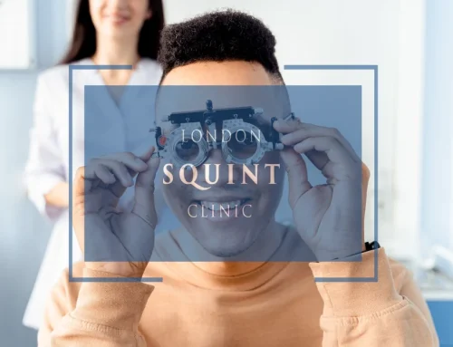
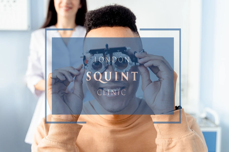
In the UK, squint surgery may be performed via the NHS, through insurance, or as self-pay. NHS treatment is free, but waiting times can be long and surgery is often performed by trainees under supervision. Many operations are carried out by surgeons who mainly specialise in children’s squint rather than adult complex cases.
With insurance, fees are standardised — meaning some leading specialists choose not to participate. Self-pay allows you to choose your surgeon directly and prioritise experience, specialisation, and access.
Many centres quote only a surgical fee. Hospital costs, anaesthetic fees and follow-ups are frequently additional. At London Squint Clinic, everything is included in one transparent package.
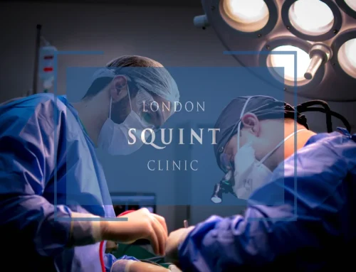
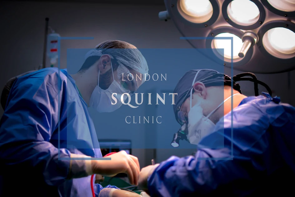
Our Complete Package – £10,000
- ✔ Advanced surgery by Mr Ali (one or both eyes)
- ✔ Adjustable sutures where clinically indicated
- ✔ Detailed orthoptic planning
- ✔ All hospital & anaesthetic fees included
- ✔ Post-operative medication
- ✔ Two video follow-ups
- ✔ Face-to-face review appointment
What Makes Us Different
- ✔ 100% focused on adult squint & double vision surgery
- ✔ >95% audited success rate
- ✔ Free re-treatment at 3 months if worse (extremely rare)
- ✔ 24/7 direct WhatsApp access to your surgeon during recovery
- ✔ Optional well-being session & pre-op reassurance call
Initial consultation: from £150
Surgery typically within 4 weeks. No referral required. Self-pay only.
Essential Insights: Understanding and Managing Superior Oblique Palsy
- Superior oblique palsy (fourth nerve palsy) affects eye movement, causing vertical double vision and a characteristic head tilt toward the opposite shoulder to compensate.
- The condition can be congenital or acquired, with head trauma being the most common cause in adults.
- Diagnosis involves specialized tests like the Parks-Bielschowsky three-step test and sometimes neuroimaging to identify the underlying cause.
- Treatment options range from conservative approaches (prism glasses, observation) to surgical interventions that target specific muscles to restore balanced eye alignment.
- Most patients achieve good functional outcomes with appropriate treatment, though perfect alignment in all gaze positions may not always be possible.
- Seek specialist care promptly for sudden double vision, persistent head tilt, or visual symptoms following head trauma.
Table of Contents
- Understanding Superior Oblique Palsy: Causes and Symptoms
- The Fourth Cranial Nerve: Anatomy and Function Explained
- Why Do Patients Tilt Their Head with Superior Oblique Palsy?
- Diagnosing Trochlear Nerve Palsy: Tests and Assessments
- Treatment Options for Superior Oblique Muscle Weakness
- Surgical Approaches to Correct Fourth Nerve Palsy
- Living with Superior Oblique Palsy: Management Strategies
- When Should You See a Specialist for Head Tilt Diplopia?
Understanding Superior Oblique Palsy: Causes and Symptoms
Superior oblique palsy, also known as fourth nerve palsy or trochlear nerve palsy, is a condition affecting one of the six muscles that control eye movement. This specific muscle, the superior oblique, is responsible for moving the eye downward and outward. When the fourth cranial nerve that supplies this muscle becomes damaged or dysfunctional, patients experience characteristic symptoms that significantly impact their vision and daily life.
The causes of superior oblique palsy can be broadly categorised as either congenital (present from birth) or acquired. Congenital fourth nerve palsy may result from developmental abnormalities or birth trauma. Acquired causes include:
- Head trauma (the most common cause in adults)
- Vascular disorders such as diabetes or hypertension
- Tumours or aneurysms compressing the nerve
- Viral infections
- Multiple sclerosis
- Idiopathic causes (where no specific cause can be identified)
The primary symptoms of superior oblique palsy include vertical diplopia (seeing two images stacked one above the other), which typically worsens when looking down and to the opposite side of the affected eye. Patients often develop a compensatory head tilt toward the opposite shoulder to maintain single vision. This characteristic head position is a key diagnostic feature that ophthalmologists look for when assessing potential fourth nerve weakness.
The Fourth Cranial Nerve: Anatomy and Function Explained
The trochlear nerve, or fourth cranial nerve, is the smallest of the twelve cranial nerves but has the longest intracranial course. This unique nerve originates from the dorsal midbrain, crosses to the opposite side (decussates), and exits the brainstem dorsally—making it the only cranial nerve to emerge from the back of the brainstem. This complex anatomical pathway makes the fourth nerve particularly vulnerable to injury, especially during head trauma.
The primary function of the trochlear nerve is to innervate the superior oblique muscle of the eye. This muscle passes through a fibrocartilaginous pulley called the trochlea (from which the nerve derives its name) before attaching to the eyeball. When functioning correctly, the superior oblique muscle performs two critical actions:
- Intorsion (rotating the top of the eye toward the nose)
- Depression (moving the eye downward, particularly when the eye is adducted or turned inward)
The fourth cranial nerve is unique among the ocular motor nerves in that it innervates the contralateral superior oblique muscle. This means that the right trochlear nerve controls the left superior oblique muscle and vice versa. This anatomical arrangement explains why damage to one side of the brain can affect the opposite eye’s movement.
Understanding this complex anatomy helps explain why patients with superior oblique palsy experience specific patterns of double vision and why they adopt characteristic head positions to compensate for their visual disturbance. The intricate pathway of the fourth nerve also explains why it is particularly susceptible to damage during traumatic brain injuries.
Why Do Patients Tilt Their Head with Superior Oblique Palsy?
The characteristic head tilt observed in patients with superior oblique palsy is not merely a random posture but a deliberate compensatory mechanism to maintain single vision. This adaptive behaviour provides valuable diagnostic clues for clinicians and represents the body’s remarkable ability to adjust to visual disturbances.
When the superior oblique muscle is weakened, it cannot effectively perform its primary functions of intorsion and depression of the eye. This weakness creates a vertical misalignment between the eyes, resulting in troublesome double vision (vertical diplopia). The brain, seeking to restore single vision, instinctively guides the patient to tilt their head toward the shoulder opposite to the affected eye.
This compensatory head position works through several mechanisms:
- By tilting the head, the patient effectively rotates their visual world, minimising the vertical separation between the two images
- The head tilt triggers ocular counter-rolling reflexes that partially compensate for the muscle weakness
- This position places less demand on the weakened superior oblique muscle
For example, a patient with right superior oblique palsy typically tilts their head toward the left shoulder. This head tilt becomes more pronounced when looking down, such as when reading or navigating stairs, as these activities require more action from the superior oblique muscle.
Many patients with long-standing superior oblique palsy develop chronic neck discomfort due to this persistent head tilt. Some may be unaware of their compensatory posture until it’s pointed out by others or identified during a clinical examination. At London Squint Clinic, we carefully assess these postural adaptations as part of our comprehensive evaluation of extraocular muscle dysfunction.
Diagnosing Trochlear Nerve Palsy: Tests and Assessments
Accurate diagnosis of trochlear nerve palsy requires a systematic approach combining clinical observation, specialised tests, and sometimes advanced imaging. The diagnostic process typically begins with a thorough history-taking, focusing on the onset of symptoms, presence of head trauma, and any associated neurological complaints.
The clinical examination includes several key components:
- Head posture assessment: Observing the characteristic head tilt toward the shoulder opposite to the affected eye
- Cover-uncover test: Revealing vertical misalignment of the eyes
- Three-step test (Parks-Bielschowsky test): A cardinal diagnostic procedure that systematically identifies which extraocular muscle is dysfunctional
- Double Maddox rod test: Measuring the degree of cyclotorsion (rotation) of the eyes
- Prism cover test: Quantifying the magnitude of the vertical deviation in different gaze positions
For acquired fourth nerve palsy, particularly when associated with trauma or when other neurological symptoms are present, neuroimaging may be necessary. MRI scans can identify structural abnormalities affecting the trochlear nerve, while high-resolution MRI with thin cuts through the brainstem may visualise the nerve itself.
Distinguishing between congenital and acquired superior oblique palsy is crucial for treatment planning. Congenital cases typically show:
- Large vertical fusional amplitudes (the ability to fuse images despite misalignment)
- Facial asymmetry due to long-standing head tilt
- Minimal symptoms despite significant objective findings
In contrast, acquired cases usually present with more symptomatic diplopia, normal facial symmetry, and limited ability to fuse the double images. These diagnostic nuances guide the ophthalmologist in determining the most appropriate treatment approach for each patient.
Treatment Options for Superior Oblique Muscle Weakness
The management of superior oblique palsy encompasses a range of non-surgical and surgical interventions, tailored to the severity of symptoms, duration of the condition, and individual patient factors. Treatment aims to eliminate double vision, correct abnormal head posture, and restore comfortable binocular vision.
Non-surgical options are typically considered first, especially for mild cases or recently acquired palsies that may resolve spontaneously:
- Observation: Newly acquired fourth nerve palsies, particularly those following head trauma, may improve over 6-12 months without intervention
- Prism glasses: Fresnel prisms or ground-in prisms can compensate for vertical misalignment, providing immediate relief from diplopia
- Occlusion therapy: Patching one eye or using frosted lenses eliminates double vision but sacrifices binocular vision
- Botulinum toxin injections: Temporary weakening of the antagonist muscle can balance the forces acting on the eye and may be useful as a diagnostic tool before permanent surgical correction
For persistent symptomatic superior oblique palsy, surgical intervention offers more definitive correction. The timing of surgery depends on several factors, including:
- Stability of measurements over at least 6 months
- Severity of symptoms and impact on quality of life
- Presence of significant compensatory head tilt
- Failure of conservative measures to provide adequate relief
The specific surgical approach is determined by the pattern and magnitude of the vertical deviation, the presence of torsional components, and whether the condition is unilateral or bilateral. Modern surgical techniques can address both the vertical misalignment and the torsional component of superior oblique palsy, providing comprehensive correction of this complex disorder.
Surgical Approaches to Correct Fourth Nerve Palsy
Surgical correction of fourth nerve palsy requires a tailored approach based on the specific pattern of muscle dysfunction, the magnitude of deviation, and the presence of torsional components. The goal is to restore balanced ocular alignment in all directions of gaze, eliminate diplopia, and correct abnormal head posture.
Several surgical strategies may be employed, often in combination:
- Inferior oblique weakening procedures: Since the inferior oblique muscle acts as an antagonist to the superior oblique, weakening it can help balance the forces acting on the eye. Techniques include myectomy (removing a segment of the muscle), recession (repositioning the muscle), or anteriorisation (changing the muscle’s vector of pull)
- Superior oblique tuck or resection: For cases with lax or elongated superior oblique tendons, shortening the tendon can enhance the muscle’s function
- Vertical rectus muscle surgery: Adjusting the inferior rectus muscle of the affected eye or the superior rectus of the contralateral eye can address vertical misalignment
- Harada-Ito procedure: This specialised technique targets the anterior fibres of the superior oblique tendon to correct torsional diplopia
- Adjustable suture techniques: These allow fine-tuning of muscle position in the immediate postoperative period to optimise alignment
For complex or large-angle deviations, a combined approach involving multiple muscles may be necessary. The surgical plan must account for both primary position alignment and incomitant deviations (those that vary in different gaze positions).
Outcomes of surgical correction for superior oblique palsy are generally favourable, with success rates of 80-90% for achieving functional alignment in primary position and downgaze. However, patients should understand that perfect alignment in all gaze positions may not be achievable with a single procedure, and some may require additional interventions to address residual diplopia in secondary positions of gaze.
Living with Superior Oblique Palsy: Management Strategies
Beyond medical and surgical interventions, patients with superior oblique palsy benefit from practical strategies to manage their condition in daily life. These approaches can significantly improve quality of life, particularly for those awaiting treatment or with residual symptoms after intervention.
Adaptive techniques for managing head tilt diplopia include:
- Ergonomic adjustments: Modifying workstations, computer screens, and reading materials to accommodate the preferred head position can reduce neck strain and improve visual comfort
- Driving considerations: Using extra caution when changing lanes or navigating junctions, as depth perception may be compromised
- Reading strategies: Tilting reading material rather than the head can reduce neck fatigue during prolonged reading
- Physical therapy: Targeted exercises to strengthen neck muscles and improve posture can alleviate discomfort from chronic head tilting
- Vision therapy: Exercises to enhance fusion and expand the range of single binocular vision
For patients with congenital fourth nerve palsy who have developed effective compensatory mechanisms, intervention may not be necessary unless the compensation breaks down due to age, fatigue, or additional visual stressors. Regular monitoring is important to ensure that compensatory mechanisms remain effective and do not lead to secondary musculoskeletal issues.
Psychological support is also valuable, as the visible head tilt and challenges with depth perception can affect self-confidence and social interactions. Support groups and counselling can help patients cope with these aspects of living with extraocular muscle dysfunction.
With appropriate management strategies and medical intervention when needed, most patients with superior oblique palsy can maintain good functional vision and lead normal, productive lives despite this challenging condition.
When Should You See a Specialist for Head Tilt Diplopia?
Recognising when to seek specialist care for symptoms related to superior oblique palsy is crucial for timely diagnosis and effective treatment. While some cases of double vision are temporary and resolve spontaneously, certain signs warrant prompt medical attention from an ophthalmologist or neuro-ophthalmologist specialising in strabismus.
You should consult a specialist if you experience:
- Sudden onset of double vision: Particularly vertical diplopia (one image above the other) that persists for more than a few hours
- Double vision following head trauma: Even minor head injuries can damage the delicate fourth cranial nerve
- Noticeable head tilt: If you or others notice that you’re tilting your head to one side to see clearly
- Worsening of double vision when looking down: Difficulty with reading, navigating stairs, or other downgaze activities
- Associated neurological symptoms: Headaches, dizziness, or other cranial nerve dysfunctions accompanying your visual symptoms
- Changes in existing head tilt patterns: For those with known superior oblique palsy, any change in symptoms requires reassessment
Frequently Asked Questions
What is the main symptom of superior oblique palsy?
The main symptom of superior oblique palsy is vertical diplopia (seeing two images stacked one above the other), which typically worsens when looking down and to the opposite side of the affected eye. Patients also commonly develop a compensatory head tilt toward the opposite shoulder to maintain single vision. This characteristic head position is a key diagnostic feature that helps identify fourth nerve weakness.
Can superior oblique palsy resolve on its own?
Yes, some cases of acquired superior oblique palsy, particularly those following head trauma, may resolve spontaneously within 6-12 months without intervention. However, congenital cases and those caused by vascular disorders or tumors typically do not resolve without treatment. Newly acquired fourth nerve palsies should be observed for several months before considering permanent surgical correction.
How is superior oblique palsy diagnosed?
Superior oblique palsy is diagnosed through a combination of clinical assessments including the three-step Parks-Bielschowsky test, cover-uncover test, double Maddox rod test, and prism cover test. Doctors also observe the characteristic head tilt and evaluate eye movement patterns. In some cases, MRI imaging may be necessary to identify structural abnormalities affecting the trochlear nerve, particularly for acquired cases with neurological symptoms.
What causes superior oblique palsy?
Superior oblique palsy can be congenital (present from birth) or acquired. Common causes include:
– Head trauma (most common in adults)
– Vascular disorders such as diabetes or hypertension
– Tumors or aneurysms compressing the nerve
– Viral infections
– Multiple sclerosis
– Idiopathic causes (no identifiable cause)
Congenital cases may result from developmental abnormalities or birth trauma.
What is the most effective treatment for superior oblique palsy?
The most effective treatment for superior oblique palsy depends on the severity and cause. Non-surgical options include prism glasses, occlusion therapy, and botulinum toxin injections. For persistent symptomatic cases, surgical intervention offers more definitive correction, with success rates of 80-90%. Surgical approaches may include inferior oblique weakening procedures, superior oblique tuck or resection, vertical rectus muscle surgery, or the Harada-Ito procedure for torsional diplopia.
Why do patients with superior oblique palsy tilt their head?
Patients with superior oblique palsy tilt their head toward the shoulder opposite to the affected eye as a compensatory mechanism to maintain single vision. This head tilt works by rotating their visual world to minimize vertical separation between images, triggering ocular counter-rolling reflexes that partially compensate for muscle weakness, and placing less demand on the weakened superior oblique muscle. This instinctive adaptation helps the brain restore single vision despite the muscle dysfunction.
Can children have superior oblique palsy?
Yes, children can have superior oblique palsy, which is often congenital in this age group. Children with congenital fourth nerve palsy typically develop strong fusion mechanisms and may show fewer symptoms despite significant objective findings. They often display facial asymmetry due to long-standing head tilt. Early diagnosis and treatment are important to prevent permanent facial asymmetry and ensure proper visual development.
Related Posts


Find out if you are suitable for Double Vision Treatment
Find out if you could benefit from this life changing surgery by contacting us today
Our most popular procedures


Hello, I’m Nadeem Ali
I’m one of the few eye surgeons in the world with 100% focus on Squint and Double Vision Surgery.
I have 24 years of eye surgery experience, and worked for 13 years as a Consultant at London’s renowned Moorfields Eye Hospital.
In 2023, I left the NHS to focus fully on treating patients from across the world at the London Squint Clinic. You can read more about me here.
There’s lots of information on the website about: squint surgery, double vision surgery and our pricing.
The most rewarding part of my job is hearing patients tell me how squint or double vision surgery has changed their lives. You can hear these stories here.
Mr Nadeem Ali
MA MB BChir MRCOphth FRCSEd(Ophth)